Gallery
Images
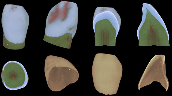
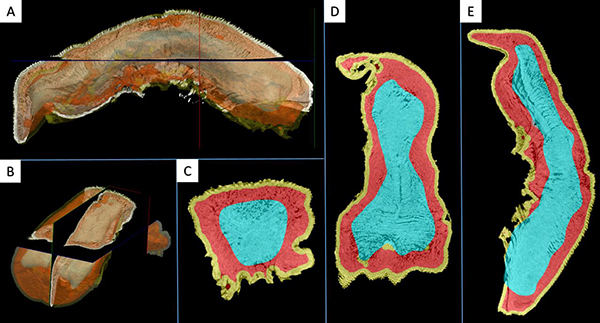
Crown and enamel in different views of the tooth
Root resorption in maxillary first molar after orthodontic force application in a mouse

Mandibular bone of a young mouse showing signs of osteoporosis
Joints
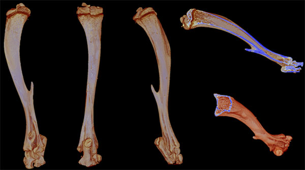
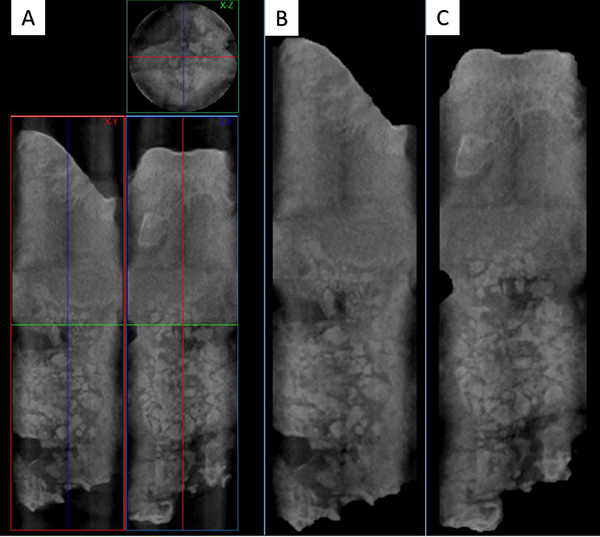
Tibia of an old mouse
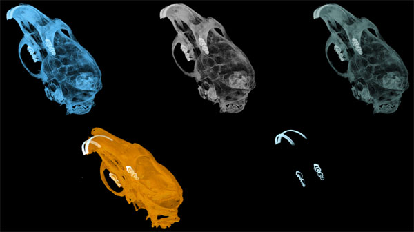
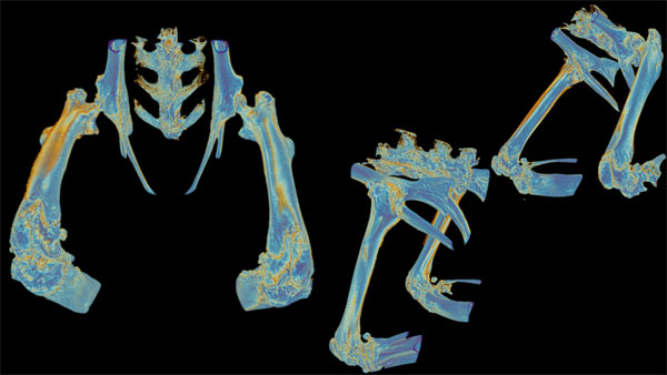
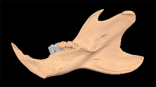
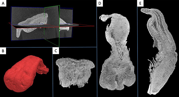
Skull and teeth of a mouse
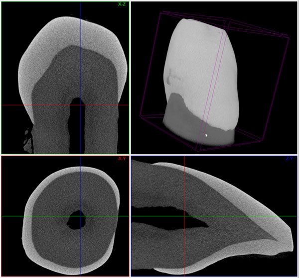
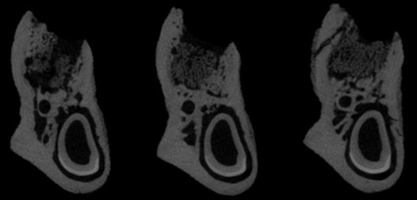
Multiplanar and 3D reconstruction of a crown of a tooth
Bone defect in tibia of an old mouse
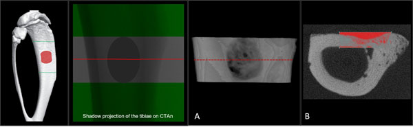
Shadow projection in the tibia of a mouse
Mandible of a rat
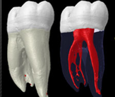
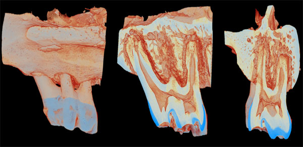
Tooth and root canal
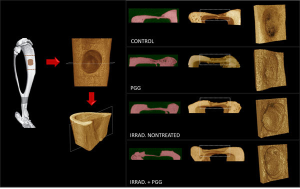
Alveolar bone repair in irradiated rat
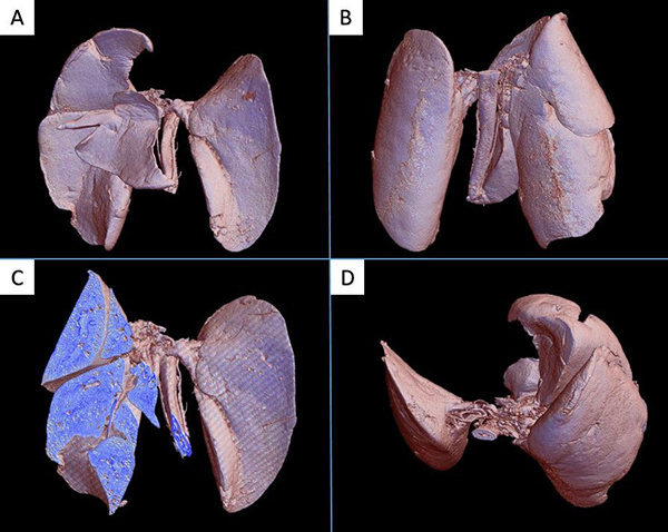
3D volume rendering reconstruction of a mice lung. A. Frontal view. B. Back view. C. Frontal view with section to show the interior of the right lung D. Top view.
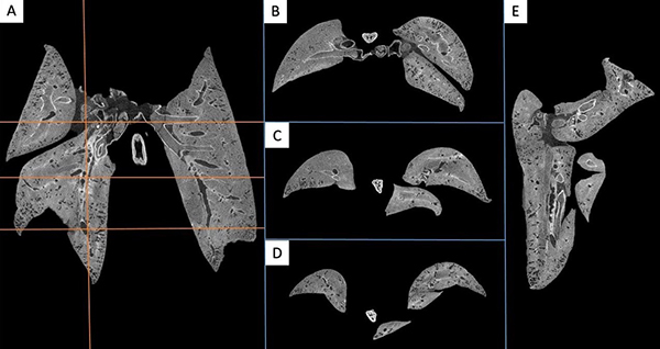
Reconstructed data of the mice lung. A. Coronal view. B, C and D. Axial view of the regions (top, middle, and bottom). E. Sagittal view of the right lung.
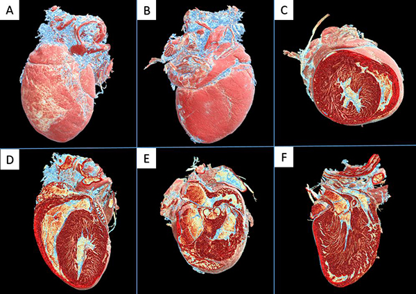
3D volume rendering reconstruction of a mice heart. A. Frontal view. B. Back view. C. Axial view of the middle. D. Coronal view of the middle front. E. Axial view of the top. F. Coronal view of the middle back.
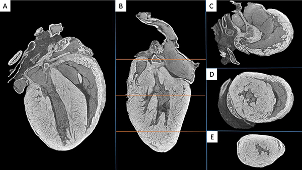
Reconstructed data of the mice heart. A. Coronal view. B. Sagittal view. C, D and E. Axial view of the regions (top, middle, and bottom).
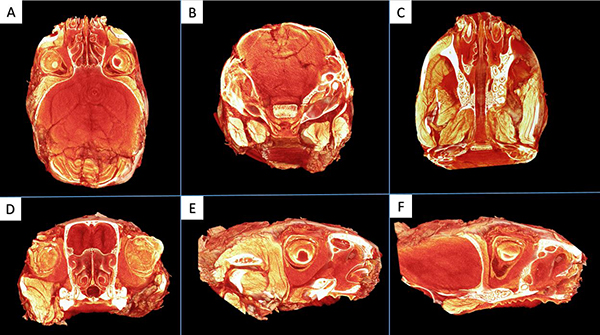
3D volume rendering of a mice head. A. Axial view of the middle. B. Coronal view of the posterior region. C. Axial view of the inferior region. D. Coronal view of the anterior region. E and F. Sagittal view.
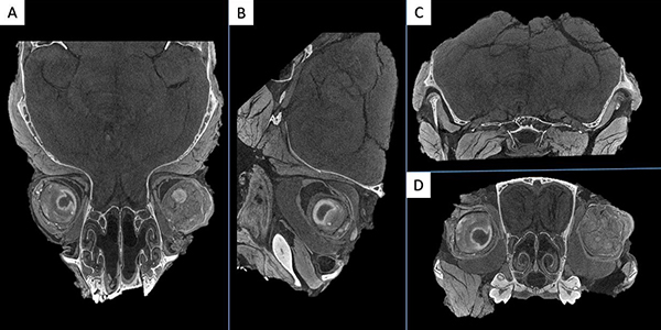
Reconstructed data of the mice head. A. Axial View. B. Sagittal view. C and D. Coronal view of the posterior and anterior region respectively.
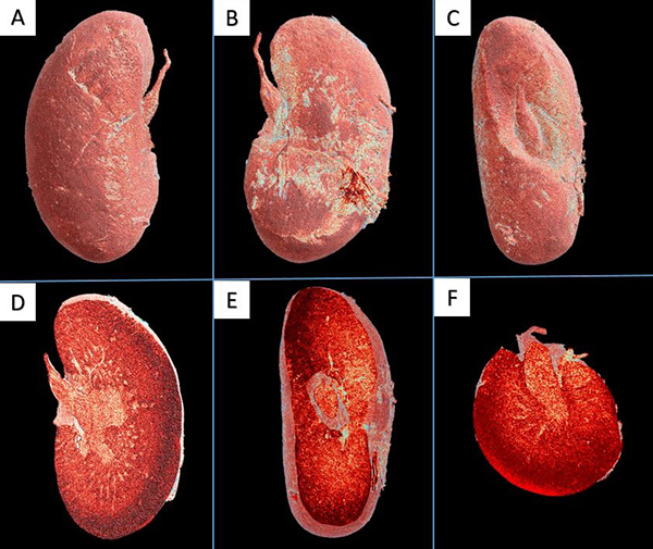
3D volume rendering of a mice kidney. A. Frontal view. B. Back view. C. Medial view. D. Coronal view. E. Sagittal view. F. Axial view.
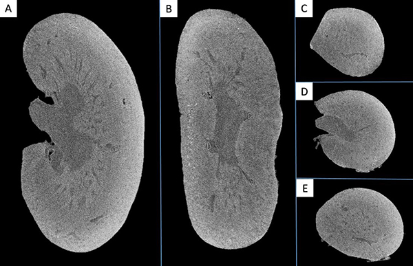
Reconstructed data of the mice kidney. A. Coronal view. B. Sagittal view. C, D and E. Axial view of the regions (top, middle, and bottom).
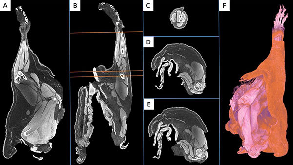
Reconstructed data of a mice arm. A. Coronal view. B. Sagittal view. C, D and E. Axial view of the regions (top, middle, and bottom). F. 3D volume rendering.
A and B. 3D Volume rendering of a mice tongue. C, D, and E. Reconstructed data of a mice tongue (coronal, axial and sagittal view) with the segmentation of the different anatomic structures.
A. Reconstructed data showing the three planes. B. 3D volume rendering of a mice tongue. C, D, and E. Reconstructed data of a mice tongue (coronal, axial and sagittal view).
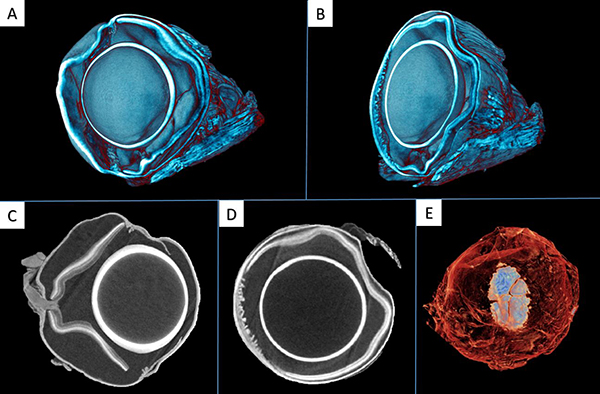
A and B. 3D volumen rendering reconstruction of a eye mice. C and D. Reconstructed data sagittal and coronal view. E. 3D volumen rendering reconstruction frontal view.

3D volume rendering reconstruction of a bracket.
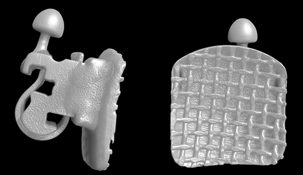
3D volume rendering reconstruction of a bracket.
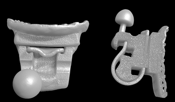
3D volume rendering reconstruction of a bracket.
Bone graft. A. Reconstructed data in the three planes. B and C. Sagittal and coronal view of a bone graft
Videos

Crown Example

Tibia Example

Skull Example

Lung Example

Bracket Example

Teeth Example
Storm-dominated diatomite: transport and deposition by their micro-texture