The Loma Linda University School of Dentistry was well represented at the week long 64th Annual Session of The American Academy of Oral and Maxillofacial Radiology (AAOMR) held recently at the Beverly Hilton Hotel in Beverly Hills, California.

recognition lunch.
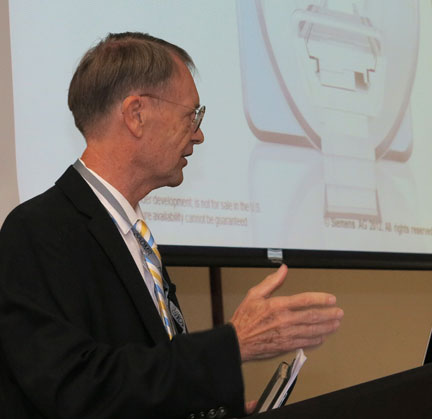
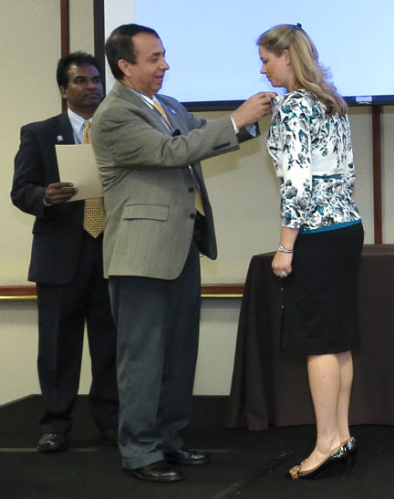
Edwin L. Christiansen, DDS’75A, PhD, professor, School of Dentistry, adjunct professor, Department of Radiology, School of Medicine, was the invited speaker for the ABOMR Recognition Lunch and Oration ceremony at which Heidi Kohltfarber, DDS’03, MS’12, assistant professor, Oral Diagnosis, Radiology, and Pathology, was inducted as a diplomate, having successfully challenged the board’s rigorous certification examination process.

Attending the ceremony and witnessing Dr. Kohltfarber’s induction were Ronald Dailey, PhD, dean, LLUSD, Heidi Christensen, DDS’83, MS, chair, Department of Oral Diagnosis, Radiology and Pathology, Kenneth Abramovitch, DDS, MS, professor, Department of Oral Diagnosis, Radiology and Pathology, director, Radiology and Imaging Services, and president-elect of the ABOMR, and Dwight Rice, DDS’96, Department of Oral Diagnosis, Radiology and Pathology. Dr. Kohltfarber will be returning to LLUSD in July of 2014 from study leave at the University of North Carolina.
Dr. Christiansen’s address at the ABOMR Recognition Lunch and Oration was entitled, “BCT ET cet.” He underscored the accomplishment of the AAOMR for its persistence in urging advanced training in the science and art of maxillofacial radiology in dentistry and its importance to the advancement of dental arts and sciences. He also recognized the long and productive relationship between the LLU Schools of Dentistry and Medicine through the Department of Radiology chaired by David B. Hinshaw, Jr., MD. In conclusion, Dr. Christiansen noted that the first application of advanced medical imaging (Computed Tomography) for a uniquely dental problem (the Temporomandibular joint) occurred in March of 1981; thirty-two years ago.
Three research studies representing collaboration between LLUSD personnel, the LLU School of Medicine faculty and other researchers were presented during the week by LLUSD researchers.
Effect of CBCT Training Sessions in Detecting Apical Bone Defects
Rice D, Luikham V, Christensen H, Torabinejad M, Oyoyo U.
There are no evidence-based guidelines for training to determine competency in identifying pathosis with CBCT. As a consequence, this study measured the effect that multiple small group and individual CBCT training sessions would have on the ability of fourth year dental students to interpret artificially created periapical lesions in human mandibles.
Using receiver operating characteristics (ROC) analysis, post training detection rates more accurately predicted the presence of lesions at a statistically significant level.
The Asymmetry Index (AI)
Abramovitch K, Huynh CP, Owen H, Bailey JR, Zhang WZ, Spira M.
This study was sparked by the absence of a standardized methodology for measuring skull symmetry, despite the fact that CBCT provides excellent 2D and 3D anatomic visualization.
Using CBCT scans of 15 Caucasian females (ages 19-29), this preliminary study showed that 3D-AF measurements are reliably reproducible, with mandibular structures (i.e., lingual foramen, mental foramen, lateral condylar pole) demonstrating the greatest degree of facial asymmetry (AI>3.0).
Study findings suggest an asymmetry index of 3.0 as a threshold for notable asymmetry, and mandibular structures demonstrate more asymmetry than maxillary structures. Replication of this study on a larger sample size has the potential to validate the current findings.
Evaluation of Bacterial Contamination on Photostimulable Phosphor Plates
Christensen H, Rice D, Cross J, Ordelheide E, Roquiz D, Aprecio R.
This study set out to discover whether bacterial cross-contamination occurs between patients when PSP places and barrier envelopes are used as directed in the pre-doctoral and dental hygiene clinics at Loma Linda University School of Dentistry.
Based on the study, barriers appear to adequately prevent PSP-plate cross-contamination between patients. However, the possibility of cross-contamination is still a concern; so universal precautions and strict adherence to radiology infection control procedures are essential for protecting our patients.
