LLUSD dental students from the class of 2011, and dental hygiene students from the class of 2010, made their Student Research Presentations as computerized slide shows at Loma Linda University’s Centennial Complex on March 18.
The twelve winning teams of presenters, divided among three categories (laboratory,
clinical, and
educational), are pictured below, accompanied by the abstracts that describe their research.
Laboratory 1st Place
Changes in Shade and Glossiness Relative to Changes in pH Found in Bleaching Agents
STUDENTS:Joohee Oh, Sang Yoo
MENTOR: Dr. Sean Lee
Objective: To test the effectiveness of tooth whitening agents on the shade and gloss of teeth by using 35-38% H2O2 single visit in-office bleaching materials with varying pH values.
Materials and Methods: Anterior and posterior teeth (N = 100) of B2 shade or darker were carefully balanced and assigned to three treatment groups and one control group. Each of the three whitening agents was applied to teeth in their respective groups according to manufacturer’s instructions. A light was used for the application of Group 1 product only. Shade and gloss were measured using a spectrophotometer and shade measuring instrument, respectively. The Wilcoxon Signed Ranked Test was used to analyze the collected data.
Results: The results indicated that group 1 (pH=6) had the highest decrease of 2.92 in shade value, and lowest decrease of 1.36 was found in group 4 (pH=4.5). Group 2 (pH=7) had a decrease in shade value of 2.88. The gloss value in group 1 showed the highest increase of 1.27 while group 2 showed the lowest increase of 0.47. Group 4 had an increase of 0.72. Excluding the control group, all other groups showed a significant decrease in shade values (p= 0.001). Only groups 1 & 4 showed a significant increase in glossiness (p=0.026 and 0.028, respectively). There was no positive correlation between lower pH value and decrease in shade or glossiness value.
Conclusion: Based upon this in-vitro study, high concentrations of H2O2 do not adversely affect the glossiness of the teeth. The effects of pH level on shade or glossiness were insignificant. Clinical Implications: Lower pH concentration does not decrease glossiness nor increase the whitening effect of in-office tooth bleaching agents.
Laboratory 2nd Place
Electromechanical Luxation: An Application of Dynamic Loading
STUDENTS: Larina Chu, Bryce Chun, Michael Hiersche
MENTORS: Dr. Alan Herford, Dr. Mei Lu

Michael Hiersche, Larina Chu, Bryce Chun
Objectives: The electromechanical luxation (EML) technique applies dynamic loading to a tooth through use of an electromechanical actuator. Compared with traditional extraction, this study was to demonstrate the advantages of the application of EML that include the ability to luxate dentition through use of minimal force, decreased time required for extraction post dynamic loading, and reduced trauma during extraction.
Methods: Three pig heads were used with each mandible and maxilla serving as experimental group on one side and control group on the other side. Total of forty-four premolars and molars were extracted. Twenty-two teeth in the control group were extracted by traditional extraction technique using elevators and forceps, while twenty-two teeth in the experimental group were luxated by EML technique and then extracted with proper elevators and forceps. The EML technique utilized an electromechanical actuator to luxate the pig dentition. A load cell measured the applied force during dynamic loading. Tooth mobility was measured pre- and post-EML. The time of extraction was recorded for each tooth. Post extraction, the number of tooth fragments was documented to compare trauma during extraction.
Results: After luxation, the EML technique demonstrated a statistically significant reduction in extraction time. The experimental group demonstrated a statistically significant increase in mobility post dynamic loading with an average of 1.9mm. The number of documented tooth fragments was less for pig molars with the EML technique. However, no reduction in tooth fragments for the premolars was noted. Maximum of 100 psi was applied to luxate the dentition.
Conclusion: EML can be used to luxate teeth by increasing mobility and reducing extraction time with minimal force. This technique also indicates reduced trauma during extraction. This technique warrants a clinical application in oral and maxillofacial surgery, especially in cases of limited open jaw space and/or compromised coronal structure.
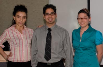
Laboratory 3rd Place
In-vitro Comparison of Various Torque Drivers in Implant Prosthodontics
STUDENTS: Jeri Bullock, Christy Pogue, Sophia Sellas
MENTOR: Dr. Jaime Lozada
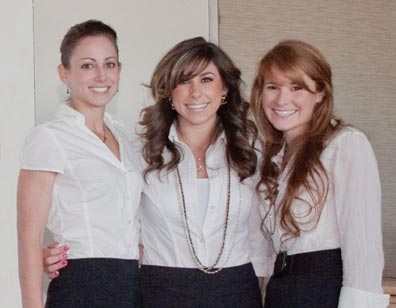
Jeri Bullock, Sophia Sellas, Christie Pogue
Objective: A major cause of prosthodontic implant failure is inconsistencies in proper calibration of mechanical torque limiting devices (MTLDs) leading to either over tightening or under tightening of dental implants. The purpose of this study is to determine any inconsistencies in calibration of MTLDs after multiple use and sterilization.
Methods: Two groups each containing four different MTLDs were tested by three experienced operators; Group 1 included one friction style and three spring style devices new from the manufacturer (Control Group). Group 2 included the same four MTLDs that have been subjected to multiple use and sterilization. Each MTLD was tested ten times per operator, the used and sterilized MTLDs were then sterilized twenty-eight times and re-tested ten more times per operator. To measure the output of each MTLD a digital torque tester with a 3-jaw chuck to hold the driver was used. The data was statistically analyzed using ANOVA.
Results: There is not a statistically significant difference in the percent error between the experimental measurements and the accepted values within each system of new and used MTLDs (F=7.90, p=0.107). There is a statistically significant difference in the percent error between the types of MTLD systems (F=13.715, p=0.004). More specifically, all brands are significantly different from each other (p<0.001) in the following order of percent error: Nobel Biocare Active (mean =-0.010, std. error = 0.003), Nobel Biocare Replace (mean=0.026, std. error=0.004), Straumann (mean=-0.051, std. error=0.004), and Astra (mean=-0.081, std. error=0.004). No interaction effects are statistically significant.
Conclusion: Considering the limitations of this study, used and sterilized MTLDs show no statistically significant difference, but a clinical significance still exists according to the literature reviewed. Further studies with larger sample sizes may reveal a statistical significance. The study did show statistical significance in the overall variance between MTLD systems.
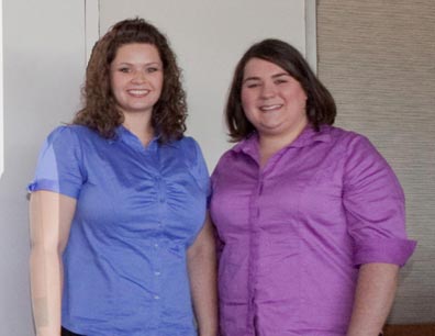
Laboratory, Dental Hygiene, 1st Place
Uptake of Fluoride Foam: A Laboratory Study on Human Enamel
STUDENTS: Lauren Stewart, Stacy Stroup
MENTORS: Dr. Wu Zhang, Ms. Joni Stephens
Stacy Stroup, Lauren Stewart
Purpose: The purpose was to determine the effectiveness of topical acidulated phosphate fluoride (APF) foam when compared to the clinically proven effectiveness of topical acidulated phosphate fluoride gel.
Materials and Methods: The method was similar to that of FDA test method #40 for fluoride uptake by enamel. Test products were an APF gel and APF foam from the same manufacturer and each contained 1.23% fluoride ion. Human 3rd molars were sectioned into buccal and lingual halves to get a total of 30 samples. Each sample was randomly assigned to a control, foam, or gel group. One-quarter inch working surfaces were isolated from each sample. Fluoride-rich outer layer of enamel was removed and baseline readings were found using fluoride electrode. White spot lesions were induced and treatment with gel or foam was performed for both one and four minutes, following manufacturer’s guidelines. Analysis was performed and compared against fluoride standard curve.
Results: After 4 minutes, mean fluoride uptake showed control = 0.19 ± 0.08, foam = 3.90 ± 3.68, gel = 9.57 ± 5.18. After 1 minute, mean fluoride uptake showed control = 0.33 ± 0.18, foam = 2.03 ± 0.91, gel = 5.82 ± 1.56. Statistical analysis showed significantly that Gel> Foam> Baseline at p=0.005 for both exposure times.
Conclusion: The results demonstrated that gel had significantly higher mean fluoride uptake into the enamel at both time intervals when compared to the foam
Clinical 1st Place
The Association Between Caries Risk Assessment Levels and Streptococcus mutans and Lactobacillus levels
STUDENTS: Adam Burr, Michael Gilman, Patrick Hachee
MENTOR: Dr. Brian Novy

Patrick Hachee, Adam Burr, Michael Gilman
Objective: It has been assumed that a high Caries Risk Assessment level indicates that a patient has dental caries, which is ultimately diagnosed by high intraoral bacterial levels. We are attempting to verify the assumption that a high CRA level does in fact correlate with a high intraoral bacterial load of Lactobacillus and Streptococcus mutans.
Methods: Subject caries risk levels and intraoral bacteria and ATP
levels were previously determined during a student cariology course. Subjects were either classified as high or low caries risk depending on responses to inquiries on LLUSD clinical Caries Risk Assessment form. Two intraoral bacterial tests were performed on each subject. The first used a saliva sample to grow a culture on a cariogenic bacteria selective plate. The other tested intraoral measured biofilm ATP levels. We will take a retrospective look at whether or not there is a statistically significant association between assessed caries risk levels and actual intraoral bacteria and ATP levels.
Results: A Chi-Square analysis found a significant association between high and low CRA levels and high and S. mutans and Lactobacillus levels, respectively. There are no significant differences between CRA levels and mean ATP RLUs. However there are significant differences between S. mutans and Lactobacillus levels and mean ATP RLUs.
Conclusion: Patients who are classified as High Risk or Low Risk on CRA are more likely to have high or low amounts of the cariogenic bacteria S. mutans and Lactobacillus, respectively. CRA levels do not associate with respective ATP RLUs. But ATP measurements do significantly identify the presence of cariogenic bacteria. The CRA form used at LLUSD is indeed helpful in the individualized treatment and prevention of caries.
Clinical 2nd Place
Two or More Canals in Mandibular Bicuspids
STUDENTS: Christina Chun, Joanne Oh
MENTOR: Dr. David Jaramillo

Joanne Oh, Christina Chun
Objective: To find the incidence of canal variation in mandibular premolars, such as multiple and bifurcated canals, using cone beam tomography.
Introduction: Lack of attention to canal morphology leads to inadequate cleaning and shaping during root canal treatment leading to clinical failures.
Literature review: Of the 4,733 mandibular first premolar studied by previous researchers, 24.2% had a more than one canal, and of the 3,063 mandibular second premolars studied, 9% had more than one canal.
Method: Analyze and record number of canals in mandibular premolars of fifty cone beam patient scans from a saggital and horizontal point of view in 0.04mm increments.
Results: Of the 100 first premolars, 33% had more than one canal, and of the 100 second premolars, 30% had more than one canal.
Conclusion: The incidence of multiple canalled mandibular premolars is higher than previously recorded results, especially the second mandibular premolars. However results may be subject to error due to variable human judgment when analyzing the cone beam scans. Never the less, the results indicate that there is a wide variability in canal morphology in mandibular premolars. Therefore clinicians should be more aware of the incidence of multiple canals when providing root canal therapy.
Clinical 3rd Place
SEM Examination of Restoration Margins in Vivo: An Observational Study
STUDENTS: Dane Andersen, Ray Arsenault, Justin Schmidt
MENTOR: Dr. Brian Novy
Justin Schmidt, Dane Andersen, Ray Arsenault
Objective: In our study we devised a protocol and procedure for a closer examination of restorations in-vivo. The focus of our research examined the margin interface between composite, GI, and amalgam restorations in natural teeth under SEM using a highly sensitive impression material. This study explored the viability of studying impression materials using a scanning electron microscope to obtain pictures of widely used dental materials and marginal restoration breakdown.
Methods: We took alginate full-arch impressions with stock trays. The alginate impression was poured up in microstone or plaster to make a full-arch cast. A custom tray was then made of the cast arch. The custom tray lacks retention features for the polyether impression. Next, a full-arch impression was taken using a polyether material with the custom tray. The custom tray was then removed and separated from the polyether impression material. The impression material was trimmed with a #25 blade to allow the impression to sit as flat as possible. The impression was again trimmed down to a straight line at or slightly above the gingival margin. Then, the impressions were allowed to sit for at least 24 hours in a desiccator. After they have been dried completely, they were then viewed under a scanning electron microscope. The restorations were then examined using the impression, intra-oral photographs, and scanning electron microscope images.
Results: Images obtained at low resolution (22x) up to high resolution (300x) with the scanning electron microscope revealed very minute details such as enamel crystals. High-resolution digital images of the impressions themselves as well as intra-oral photographs were also obtained.
Conclusions: This study demonstrates the use of a highly accurate impression material along with observation under a scanning electron microscope is an effective method for evaluating the restoration to tooth interface as well as observing natural tooth anatomy in tremendous detail.
Clinical, Dental Hygiene, 1st Place
Needle Gauge and Pain Perception
STUDENTS: Allina Lopez, Lucrecia Santana, Kristen Tayler
MENTORS: Dr. Barry Krall, Joni Stephens
Allina Lopez, Lucretia Santana, Kirsten Tayler
Purpose: To determine if needle gauge affects pain perceived while giving a local anesthesia injection. Previous research showed no correlation between needle gauge and pain perceived. Many dental professionals assume larger gauge needles cause more pain than smaller gauge needles. Most of the previous research is over 30 years old, was not standardized; injections were administered in various mouth sites, studies used local anesthetics, and different clinicians were used throughout the same study. For this study, needle insertion was standardized, penetrations were made at the same two locations in each person’s mouth without local anesthetics, and the same clinician administered all insertions.
Methods and Materials: Twenty dental and dental hygiene students were selected to receive six injections over 3 sessions within approximately three months. Three different gauge needles 25, 27, & 30 were tested. Two different needle gauges were evaluated on the right and left maxilla above the first premolar at the height of the mucogingival junction at each session. The gauge of the needle and side of penetration were randomized. Baseline vitals (HR, BP, SpO) were obtained after resting for 5 minutes comfortably. Topical was place for 1 minute before each needle was inserted 3mm into alveolar mucosa 5 mm above the mucogingival line. An unbiased observer evaluated the discomfort perceived by the participant. Both the participant and the observer were blinded to the needle gauge. The subject and the observer independently recorded on a 0 – 5 likert scale the pain level
Results: The mean score for the 25 gauge = 1.5, the 27 gauge =1.3, and the 30 gauge = 1.0. The range for the 25 G was 0-4, 27G 0-5, 30G 0-4.The mean for the non bias observer were 25 gauge .85, 27 gauge 1.1, and 30 gage 1.2. The range for 25G 0-3, 27G 0-4, and the 30G 0-3.
Conclusion: There appears there is no statistical difference in pain perceived by the patient depending on needle gauge and the pain perceived by the patients may be due to injection technique.
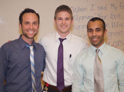
Education 1st Place
A Digital Instruction of Techniques and Principles of Mucoperiosteal Flaps
STUDENTS: Scott Arceneaux, Mike Knutson, Filip Orban
MENTORS: Dr. Mei Lu, Dr. Wayne Tanaka
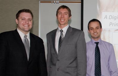
Scott Arceneaux, Michael Knutson, Filip Orban
Objective: Dental students are expected to become competent in designing and developing mucoperiosteal flaps for exodontia and preprosthetic surgeries. Because didactic mucoperiosteal flap instruction is limited to textbook illustrations and generic descriptions, dental students often struggle to comprehend and develop clinical skills related to intraoral flaps. The aim of this study was to develop and evaluate a multimedia educational resource that improves students’ clinical application and comprehension of mucoperiosteal flaps.
Methods: A multimedia computer program was created containing videos, diagrams, written instructions, and quiz questions outlining the instruments, designs, indications, and techniques of mucoperiosteal flaps commonly used for exodontia. Eighty-eight first year dental students were randomized into four evenly distributed groups based on past academic performance. Each group was then randomly assigned a study method for learning about mucoperiosteal flaps. The first group was given only a literature review. The second group used the multimedia CD in addition to the literature review. The third group used only the CD and the fourth group received no study material. After studying 0-4 hours, each student completed a clinical exam on a typodont. In addition to the clinical assessment, the students completed a multiple-choice examination. The flap learning methods were correlated with each student’s clinical and didactic performance and results analyzed.
Results: ANOVA and Scheffe analyses showed that students who trained using the multimedia CD performed better clinically and didactically than students that used only the literature review (p<.001).
Conclusion: Our multimedia CD improves student’s clinical and didactic comprehension and application of mucoperiosteal flaps. This digital instruction could help students build confidence in designing and preparing mucoperiosteal flaps and could be used widely in training dental students.
Education 2nd Place
Educational DVD for Children Ages 5-8 Years Old: Evaluating the Effectiveness of DVD as a Teaching Tool for Children Ages 5-8 Years Old As Compared to a Conventional Pamphlet
STUDENTS: Junie Baldonado, Emilynda Quinones, Michael Sacro
MENTOR: Dr. Wesley Okumura

Michael Sacro, Junie Baldonado, Emilynda Quinones
Objectives: The goals of this study are to develop an educational DVD that targets children ages 5-8 years old and their parents, and to conduct a preliminary test on the effectiveness of DVD as an educational tool versus a conventional pamphlet.
Methods: A DVD and pamphlet were developed to teach parents and children about basic concepts of tooth decay, diet, and oral hygiene. By random selection, 60 parent-child pairs will be assigned to view the DVD or to read the pamphlet. Parent-child pairs will be given a pre-test prior to viewing or reading assigned material. Parent-child pair will then be given 15 minutes to view the DVD together or read the pamphlet together. After viewing the DVD or reading the pamphlet, post-test will be administered. Results of pre-test and post-test will be compared for both DVD group and pamphlet group.
Expected results: There will be a significant improvement of post-test scores for subjects who watch the DVD form of oral hygiene education compared to post-test scores of subjects who read the conventional pamphlet form of oral hygiene education.
Conclusions: Further definitive analysis of the effectiveness of using a DVD for educating both children 5-8 years old as well as their parents is necessitated. Future study and research related to this topic is highly encouraged and may deal with several other parameters such as socioeconomic status, ethnicity, education, etc.
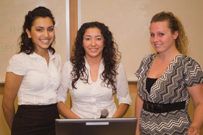
Education 3rd Place
Oral Hygiene during Orthodontic Treatment
STUDENTS: Stephanie Calvillo, Sheida Khazaii-Tabari, Isaac Penalba
MENTOR: Dr. Leroy Leggitt
Sheida Khazaii-Tabari, Isaac Penalba, Stephanie Calvillo
Objective: Create a convenient, simple, and interactive tool to educate orthodontic patients about the importance of good oral hygiene during treatment. We evaluated the efficacy of our DVD in contrast to current methods of oral hygiene instructions given by the orthodontist.
Methods: Evaluation of the DVD was conducted through a survey distributed to twenty orthodontic patients to assess their knowledge of oral hygiene before and after watching the DVD. The patients fell under the categories of LLU and non-LLU orthodontic patients. The DVD focused on the main consequences of bad oral hygiene and provided educational material on different tools and techniques to ameliorate oral hygiene during treatment.
Results: In general, the patients felt that the orthodontist did not emphasize the importance of good oral hygiene while under orthodontic treatment, 100% of the patients found the DVD to be more educational than the materials that were given to them and 91-100% of these patients would have found it helpful to have had this DVD given to them before starting orthodontic treatment.
Conclusion: There is a need to provide orthodontic patients with more practical and accessible educational material to inform them and set a baseline of knowledge for proper oral hygiene care. The DVD has proven to be efficient at accomplishing these goals and allows the patient to interactively learn about the different hygienic tools in the convenience of their own home. This can serve as an effective and marketable way of further motivating the patient in maintaining good oral hygiene during their treatment.
Education, Dental Hygiene, 1st Place
Individualized Care Case Presentation
STUDENTS: Pamela Slonaker, Hossai Tahmas, Sherwin Taylor
MENTOR: Dr. Adrian Mobilia
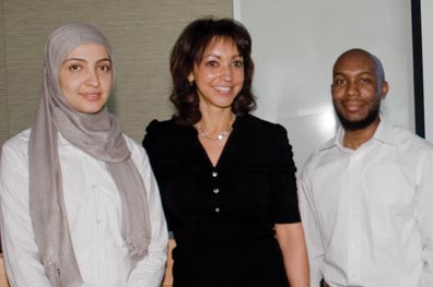
Hossai Tahmas, Pamela Slonaker, Sherwin Taylor
Objectives: The purpose of this educational case research project is to evaluate the effectiveness of a modified toothbrush, in the maintenance of oral care for the special needs patient.
Method: In this case study, a young man with cleft palate and joint disorders; arthrogryposis (stiff joints) and talepes equinovarous (club foot), was monitored over a three year span. Within the three-year span periodontal therapy was performed, oral hygiene instruction was given to the primary caregiver. Bleeding on Probing (BOP), Periodontal Pocket Depths (PPD) and Plaque score were recorded. After two years of unsuccessful change in the patient’s periodontal health, further implementation was the introduction of a modified toothbrush to the patient.
Results: Implementation of the new device and occupational therapy assistance allowed the patient to perform efficient and adequate oral care independently. There was a significant decrease in BOP and PPDs.
Conclusion: We believe that the use of this device did contribute to the patient’s improved oral health. Further periodic assessment of patient’s oral hygiene skills, and proper functionality of the device is required in each successive appointment to ensure continued efficacy.